
Clin Exp Dent Res.2021;7:786–794
Read Full Study
Abstract
Objectives: In this in vitro study, a bioluminescent marker was investigated for its potential to illuminate the assessment of dental caries and dental erosion, which are significant clinical and public health problems, through its binding of those ions, notably Ca2+, known to be released during the process of demineralization.
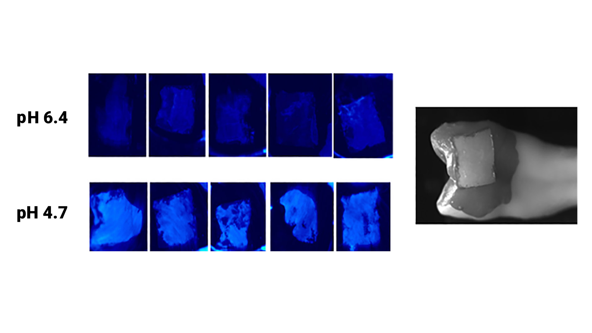
Materials and Methods: The light output from the selected bioluminescent marker was investigated in several experiments, including: (a)contact with a range of Ca2+ ion concentrations; (b) treatment of extracted teeth with solutions of differing pH, followed by application of the bioluminescence marker to assess Ca2+ ion release; and (c) application of the marker to freshly extracted teeth with natural and artificially created caries lesions on occlusal and smooth surfaces to image the Ca2+ ion distribution.
Results: The results of: experiment (a) showed that the light output from the marker increases with increasing Ca2+ concentration and of experiment (b) showed increases in light being observed as increasingly acidic solutions were applied. The results of experiment (c) showed the bioluminescence images of the extracted teeth produced “demineralization maps” of the imaged surfaces.
Conclusions: These results demonstrate the ability of a novel bioluminescence technology to image Ca2+ ions on tooth enamel surfaces which has potential in dental caries and dental erosion applications and provides the scientific basis for the ongoing development of that novel technology.