

Can you be certain of the activity status when assessing suspected early caries lesions?
A dentist or dental hygienist commonly use two methods when conducting a clinical examination of the teeth and when assessing for caries; first, is through a visual examination which is enhanced by drying the teeth along with good magnification and illumination, and second is a tactile evaluation using a probe or ball ended probe and walking that over the smooth surfaces, pits, fissures and cervical area of the teeth.
Bitewing radiographs can also show enamel caries lesions interproximally while still reversible, but they only show caries on the occlusal surface once it is into dentin.
When we detect an enamel caries lesion, the goal is to halt the caries process and reverse the lesion. The success criteria for caries prevention, therefore, should be:
- Early detection of changes in the enamel
- Confirmed identification of the activity status of a suspected caries lesion
- Simple and effective communication of the seriousness of the suspected lesion
to the patient, to encourage a behavioral change in their oral healthcare routine - Flexible prevention protocol that can be adapted for the individual patient

Using ICDAS allows staging of a lesion, but does not determine the activity status of a suspected demineralized caries lesion. Why does this matter?
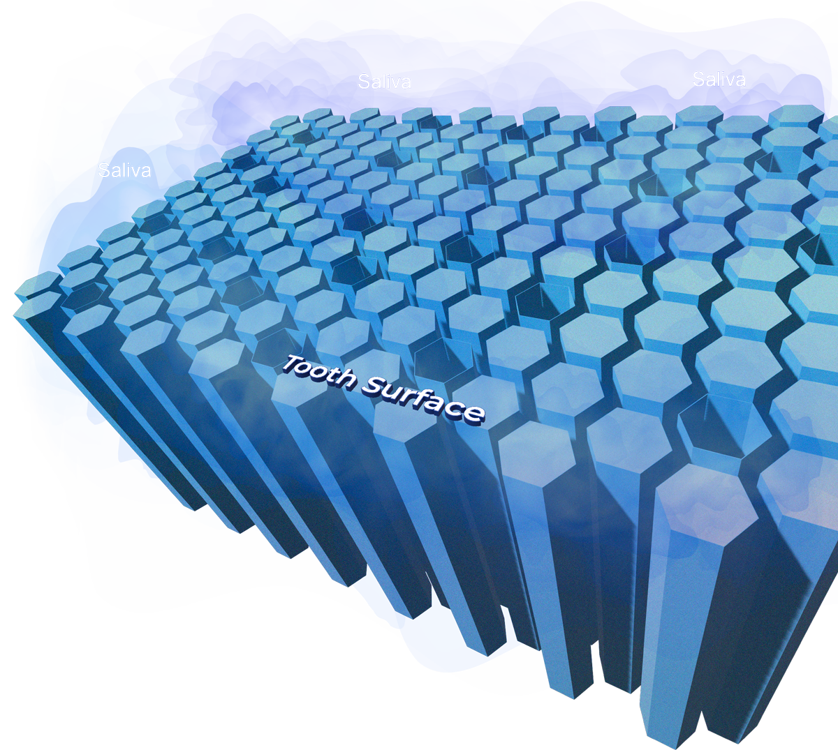
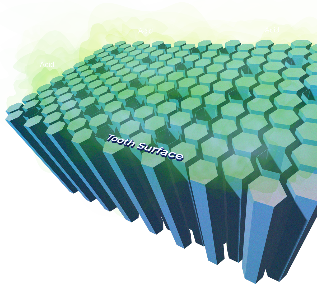
Demineralization caries activity in the enamel is characterized by the presence of free calcium and phosphate ions within the fluid-filled channels between the hydroxyapatite crystals. The calcium and phosphate ions in healthy or arrested/remineralized enamel are bound in the hydroxyapatite crystals.
What is Demineralization?
Demineralization happens when the mineral content on your teeth begins to wear away.
Can teeth Remineralize?
If caught early, demineralization can be reversed with an effective treatment plan.
Phosphate
In a caries lesion, the free calcium and phosphate ions can present as a visible change to the enamel, commonly known as a white spot lesion. When we see a white spot lesion which has a very chalky appearance and feels ‘less smooth’ when running a probe over it, we normally conclude that there is caries activity.
- Sometimes that lesion is stained and the challenge in this instance is knowing whether that is arrested or active, particularly in the fissures.
- Another part of the tooth where there may be caries activity that is challenging to identify is around the margins of restorations and fissure sealants.
- So, in instances of visible chalky white rough-textured lesions, most clinicians will conclude the caries lesion as active and, when pieced together with the stage of the lesion, support the creation of a preventive and/or corrective treatment plan.
- In the case of stained fissures, margins of restorations and fissure sealants, questionable developmental anomalies vs active lesions conclude that there is a need for more information to piece the caries diagnostic jigsaw puzzle together.
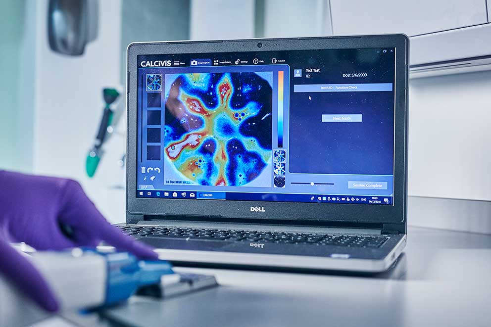
To conclude, the accuracy of whether a lesion is actively progressing or arrested/reversed, requires one to know the presence or absence of the free calcium ions in the lesion. The Calcivis Imaging System captures an image of free calcium ions and provides the missing link in caries assessments.
How does the CIS work?
The Calcivis imaging System (CIS) was developed from discovering the bioluminescent reaction of a photoprotein with calcium. Upon this discovery, a research project started to explore if this could be replicated on teeth and captured as an image, which was successful. This research then moved towards the development of a medical device that could be used chairside. This was also successful, and for the FIRST TIME EVER, clinicians can capture an image of caries activity or indeed help to determine if a lesion is arrested or reversed, thus removing the subjectivity from a visual and tactile inspection.
- Being able to capture an image of caries activity helps clinicians to decide upon the best prevention or intervention strategy.
- Being able to demonstrate no activity demonstrates that the prevention efforts from you and your patient are working.
- It is visually powerful to the patient and is a quick and simple procedure to use as part of your clinical examination.
- The CIS provides information that has never been available to any clinician. It plugs the gap in the caries assessment conundrum and is ideally placed in the hygiene room.
- It is useful for the dental hygienist in the pre-diagnosing phase of the appointment, and it is useful to the dentist for decision making; it’s a powerful communication tool to engage and motivate the patient.
The CALCIVIS® Imaging System:
- Provides information on the structure of the enamel and dentine, which can help clinicians to make an accurate diagnosis and determine the best course of treatment.
- Can detect the earliest changes in enamel and determine the activity status with clear visuals, which empower the PATIENT to make the necessary changes regarding their oral health, along with professional adjunctive therapeutics.
- Until now, there has never been a proven way to test the activity of a suspicious carious lesion.