British Dental Journal volume 231, pages 775–780 (2021)
https://www.nature.com/articles/s41415-021-3755-8#Abs1
Abstract
The CALCIVIS story is one of innovation and collaboration to deliver new technology capable of helping dentists improve patient care through solving an unmet clinical need in assessing the activity of caries lesions in enamel. Presently, there is no system routinely used in dental practice that can, in a single visit, determine whether a non-cavitated caries lesion is active or not. CALCIVIS has evolved since 2005, when a potential link between basic science in luminescence and differentiating initial-stage caries lesions that are actively demineralising and likely to progress, from other lesions which are inactive and currently do not need interventive care, was recognised. The 16-year journey has involved clinical academic dentists, scientists and entrepreneurs, general practitioners and their patients, together with serial investors and a core team working to patent, refine, assess and develop products to submit to regulatory approval and take to the international dental market. This journey has been made possible through effective long-term collaborations between disparate groups all sharing a common vision for the possibilities of harnessing new technology to help dental professionals provide better care for their patients.
Key points
- Information about caries activity in non-cavitated lesions is important to help the dental team assess the care needs of both individual tooth surfaces as well as the patient as a whole.
- Assessing caries activity or inactivity is different from merely detecting the presence or absence of lesions and staging their severity; visual activity assessments of initial-stage lesions are subjective and often unreliable.
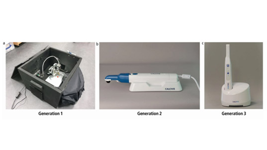
- The CALCIVIS-developed technology can, for the first time, provide an objective indication of enamel lesion activity related to the release of free calcium from demineralising lesions and a demineralisation map image can communicate the findings to both dentists and patients.